
Every breath changes the volume and pressure inside the thorax, yet the movement is difficult to infer from a motionless diagram. Airflow depends on the coordinated action of the diaphragm, chest wall, airways, and elastic lung tissue. At the same time, pulmonary arteries and veins have distinct spatial relationships with the bronchial tree that are important for understanding pulmonary circulation. A three-dimensional teaching model can give these related structures a stable spatial form, making respiratory anatomy easier to connect with the mechanics of ventilation.
Pressure Changes Behind Inhalation and Exhalation
Breathing begins with a change in thoracic volume rather than with the lungs pulling air inward by themselves. In a lung 3D model, the position of the lungs within the chest can provide the anatomical setting for explaining this pressure-based process.
When the diaphragm contracts, it moves downward and increases the volume of the thoracic cavity. Expansion of the rib cage contributes further space. The resulting drop in intrapulmonary pressure permits air to enter through the conducting airways. During quiet exhalation, relaxation of the respiratory muscles and elastic recoil reduce thoracic volume, raising pressure and moving air outward.
A rigid anatomical model cannot reproduce every movement of living tissue, but it can identify the structures responsible for the sequence. The diaphragm forms the lower boundary, the trachea leads into the bronchial system, and the paired lungs occupy distinct sides of the thorax. The mechanical explanation becomes clearer when these parts are considered together rather than as separate definitions.
Following Air Through the Bronchial Tree
The conducting pathway repeatedly divides as air travels from the trachea toward smaller bronchi and bronchioles. A three-dimensional DIGIHUMAN lung representation makes the branching pattern visible without requiring learners to assemble it mentally from several disconnected illustrations.
The right and left main bronchi enter their corresponding lungs before dividing into lobar and segmental branches. Each division reduces airway diameter while distributing air to a wider area of lung tissue. Branching is therefore both an anatomical pattern and a functional distribution system.
Rotation is useful because many branches overlap in a frontal view. An oblique or posterior perspective can reveal where one bronchus passes behind another structure. Transparent material can preserve the outline of the lungs while leaving the internal bronchial tree visible. Learners can then relate internal pathways to the organ that contains them, rather than studying the bronchial tree without the surrounding lung structure
Relating Pulmonary Vessels to the Airways
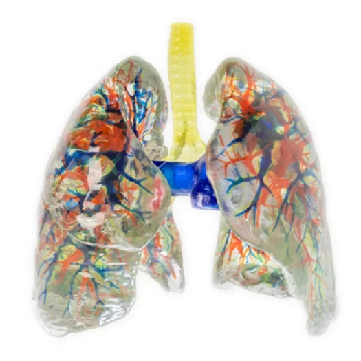
Gas exchange requires a close relationship between ventilation and blood flow. A DIGIHUMAN lung 3D model can display pulmonary arteries and pulmonary veins alongside the bronchial branches, showing how the three networks occupy the same organ while serving different functions.
Pulmonary arteries carry deoxygenated blood from the right side of the heart toward the lungs. Their branches generally accompany the airways as they extend into progressively smaller regions. Pulmonary veins return oxygenated blood to the left atrium and follow a different arrangement as they collect blood from lung tissue.
Color coding can separate these networks visually, but color should reinforce rather than replace anatomical reasoning. Learners still need to trace the direction, connections, and position of each vessel. Examining the networks from several angles also prevents an inaccurate assumption that structures lying close together always follow identical routes.
Understanding Lobes and Bronchopulmonary Segments
The lungs are not uniform masses. A segmented lung representation can distinguish the right and left lungs, their lobes, and the segmental organization associated with branches of the bronchial tree.
The right lung usually has three lobes, while the left has two and accommodates the position of the heart. Within the lobes, bronchopulmonary segments form anatomically meaningful units supplied by segmental bronchi and accompanying arterial branches. Their boundaries matter in imaging, anatomical teaching, and discussions of localized disease or surgery.
Displaying segments in different colors gives learners an initial map. The deeper objective is to understand why the divisions exist and how they relate to branching airways and vessels. A segment should not be remembered only as a colored region; it belongs to a defined route through which air and blood reach a particular part of the lung.
Turning a Physical Model Into a Teaching Sequence
Within the DIGIHUMAN product range, the professional lung 3D model uses transparent hard material so the bronchial tree, pulmonary arteries, and pulmonary veins can remain visible inside the left and right lungs. The company describes its models as being developed from high-precision digital datasets, refined segmentation information, and original tomographic scans. Individual lung segments are color-coded to assist identification.
An instructor can begin with ventilation mechanics, continue along the bronchial tree, introduce pulmonary circulation, and finish with segmental anatomy. That order links function with progressively finer structure. Another lesson may reverse the sequence by starting with a segment seen in an image and tracing its airway and vascular connections.
Physical models are most useful when handling serves an explanatory task. Learners can rotate the lungs, compare both sides, locate a branching point, and describe which structures share a region. The resulting lesson connects the pressure changes of breathing with the anatomical routes that distribute air and blood.
